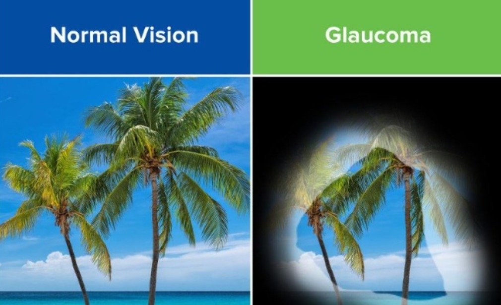
Glaucoma Services
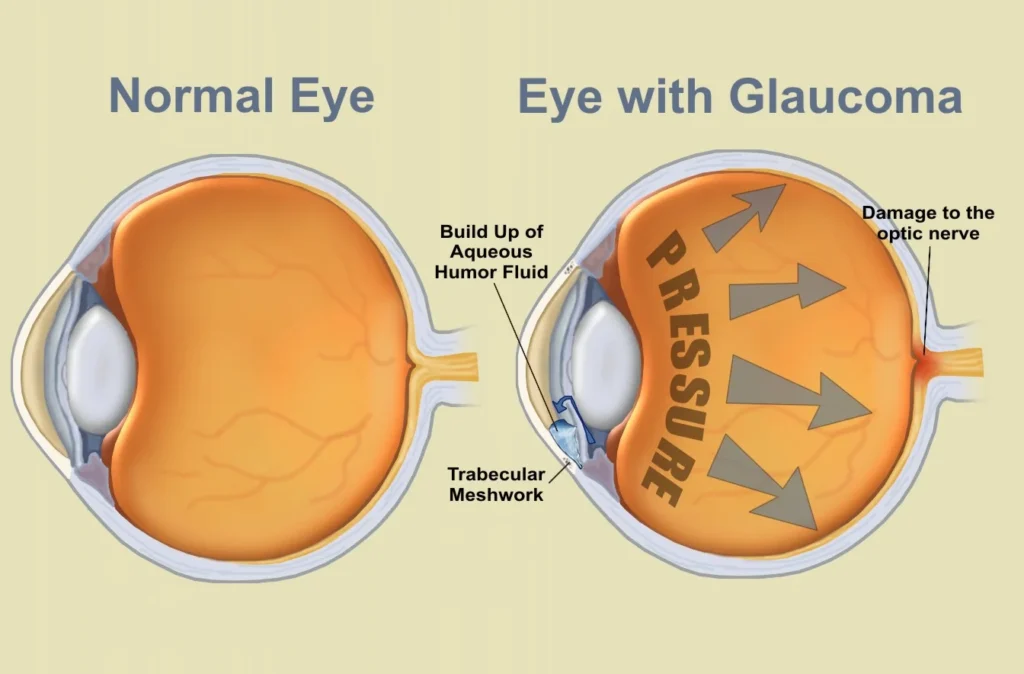
To understand glaucoma, if you can imagine that a ball is kept round with air pressure inside. And similarly the eye ball is kept round with fluid pressure inside. There is a constant ratio of inflow and out flow of a fluid called “Aqueous”. Any disturbance, either in the inflow or the outflow of this fluid will build pressure inside the eye ball. The normal pressure is 10-21mm of Hg. When this pressure raises because of the disturbance in the ratio the raised pressure inside will press on the optic nerve head. This will lead to gradual loss of peripheral vision to start with and then the central vision thus leading to total blindness. It is called a silent killer because it has no symptoms like pain for the patient to recognise the disease in time. And by the time the patient recognises the loss almost all the peripheral vision is gone and any treatment that is given is to save what is left. The only solution to this problem is to have regular examinations on yearly basis so that such a condition can be identified by the doctors. It is very frightening to see young bread winners coming in to eye clinic too late to get the treatment.
At Aggarwal Eye Hospital, the patient is investigated thoroughly with sophisticated, state-of-the-art diagnostic facilities including computerized visual field testing, optic nerve imaging, and then the treatment is personalised to the patient to maintain his vision and quality of life.
We have facilities for advanced laser and surgical procedures to control Glaucoma such as trabeculectomy, tube implants, non-penetrating glaucoma surgeries, surgery for congenital glaucoma and different lasers such as the YAG laser and Green laser.
Glaucoma Treatment in Andheri: Special Treatments We Offer
- One of the most intriguing question to today's ophthalmologist had seen the emergence of the sub-speciality of glaucoma- ‘A Silent blinder’.
- Early Clinical detection of glaucoma merely on the basis of clinical skills has been the hallmark. In-spite of the advent of newer tools clinical skills and perimetry are still the gold standards.
- Combined cataract and glaucoma surgery.
- Repeat Trabeculectomies.
- Bleb needling and revisions.
- Management of leaking/ failed blebs.
- Trabeculotomy in Paediatric age group.
- Selective Laser Trabeculoplasty (SLT)
Equipments we use for Glaucoma Surgery in Andheri.
- Goldmann Applanation Tonometer
These are standard IOLs implanted in the eye after cataract surgery for correction of distance vision only and offers improved contrast and night vision. Patients will require glasses for near vision and computer use.
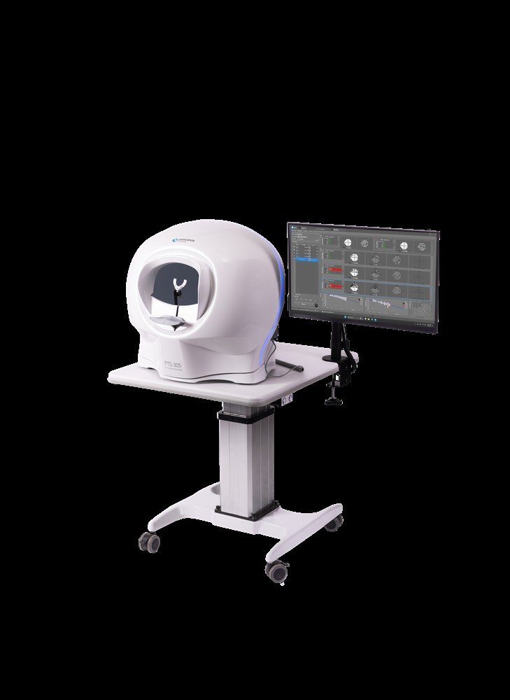
- PERIMETER OR HUMPHREY FIELD ANALYSER II
A must for glaucoma patients Instantaneous results, analysis and reports available
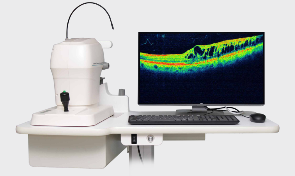
- Optical Coherence Tomography :
( OCT )is a non-invasive technology used for imaging the nerve fiber layer and to study the topography of the optic nerve. OCT, the first instrument to allow doctors to study the nerve fiber layer thickness, and is extremely useful in documenting and studying the damage due to glaucoma in its early stage before it is clinically visible. The follow up of the patients is also done very systematically and accurately with this tool.
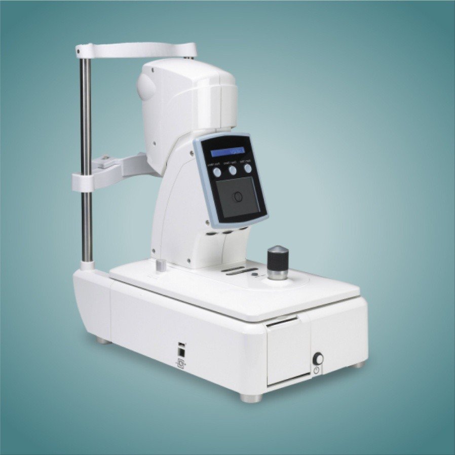
- NON CONTACT TONOMETER
It measures the intraocular pressure by optically detecting the momentary state of the cornea (applanated by air pressure) and measures intraocular pressure without touching the cornea
- PACHYMETRY
It is a simple, painless test to measure the corneal thickness. Pachymetry can help your diagnosis, because corneal thickness has the potential to influence eye pressure readings. With this measurement, your doctor can better understand your IOP reading and develop a treatment plan that is right for you.
- Ophthalmoscopy
This diagnostic procedure helps the doctor examine your optic nerve for glaucoma damage. The doctor will then use a small device with a light on the end to light and magnify the optic nerve.
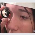
- Gonioscopy:
This diagnostic exam helps determine whether the angle where the iris meets the cornea is open and wide or narrow and closed and helps in the diagnosis of the type of glaucoma

